
Diagnosing Adenomyosis: What to Expect from Tests and Imaging
Discover how adenomyosis might be diagnosed through imaging tests. Learn what to expect and understand your symptoms better.
10 minute read
Read moreDiscover how Lotus can guide you toward lasting relief.
Explore why patients choose Lotus
Search expert-written answers, browse by topic, or find information based on where you are in your journey.
Explore All KnowledgeReach out and start your healing journey.
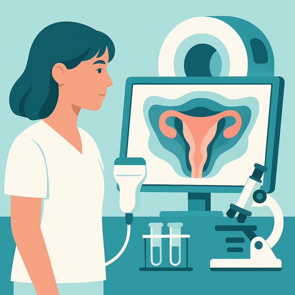
Schedule an AppointmentUnderstand how MRI and ultrasound identify adenomyosis, what to expect before and during scans, how findings are interpreted, and how results shape your treatment options.
MRI and ultrasound are the primary tools for recognizing adenomyosis and clarifying whether disease is diffuse or focal. Expert transvaginal ultrasound is often first‑line, using MUSA criteria to look for a globular uterus, heterogeneous myometrium, fan‑shaped shadowing, and small myometrial cysts. MRI adds clarity when ultrasound is inconclusive, when fibroids are also present, or before planning uterus‑sparing surgery. It highlights junctional zone changes and the extent and location of involvement, which helps distinguish adenomyosis from look‑alike conditions and informs treatment choices.
Scans are safe and radiation‑free. A pelvic MRI typically takes 30–45 minutes; contrast is seldom required for adenomyosis. Scheduling in the early to mid‑cycle can reduce false positives from normal junctional zone thickening, and antispasmodic medication may be used to limit motion. Results guide next steps—from hormonal options to conservative surgery or hysterectomy—and can shape fertility planning. For subtype‑specific guidance, see Diffuse Adenomyosis and Focal Adenomyosis; for care decisions and family‑building, see Surgical Options and Fertility Considerations. If coexisting endometriosis is suspected, complementary details are covered in MRI and Ultrasound.
“Advanced adenomyosis” usually means the adenomyosis is more extensive within the uterine muscle—often involving a larger area (diffuse disease), deeper penetration into the myometrium, and/or more pronounced changes like uterine enlargement and tenderness. It’s not the same as “advanced endometriosis,” because adenomyosis doesn’t spread outside the uterus; “advanced” is more about how much of the uterine wall appears affected and how significantly it’s impacting symptoms.
Because adenomyosis doesn’t have a single universally accepted staging system, different clinicians and radiology reports may use “advanced” to summarize imaging features (ultrasound or MRI) and the overall clinical picture—such as heavy bleeding, severe period pain, pelvic pressure, or fertility challenges. In our practice, we focus less on the label and more on what your imaging suggests (diffuse vs focal/adenomyoma, junctional zone changes, uterine size) and what your goals are (pain control, bleeding control, fertility preservation, or definitive treatment). If you’ve been told you have “advanced adenomyosis,” our team can help you interpret what that means in your specific case and map out next steps.
Endometriosis doesn’t grow at one predictable “rate.” It’s a heterogeneous condition—meaning different subtypes and lesion types can behave very differently—so one person may have slow, relatively stable disease while another has more biologically aggressive, invasive lesions that progress faster. Growth is influenced by where it is (surface vs deeper tissues or organs), the local inflammatory environment, and hormone signaling (including local estrogen activity and reduced progesterone response).
What most people notice first isn’t literal growth you can feel happening day-to-day, but changing symptoms over months or years—new bowel or bladder symptoms, worsening pain, or the appearance/enlargement of an endometrioma on imaging. It’s also why “stage” doesn’t reliably predict pain, and why a normal exam (or even normal imaging) doesn’t rule out active disease, especially with deep infiltrating endometriosis. If you’re trying to understand whether your symptoms suggest progression, our team can help you connect your symptom pattern with the most likely disease types and next diagnostic steps, and discuss when strategic excision surgery is appropriate.
A retroverted uterus (a uterus that tilts backward) is usually a normal anatomical variation, and by itself it doesn’t diagnose endometriosis. That said, endometriosis can be associated with a “fixed” or less-mobile retroverted uterus when inflammation, adhesions, or deep disease tether the uterus backward and limit how it moves on exam.
If your imaging report mentions a retroverted uterus and you also have symptoms like painful periods, deep pain with sex, bowel/bladder pain (often cyclical), or chronic pelvic pain, we look at the whole picture—not just the uterine position—to assess whether endometriosis and/or adenomyosis could be contributing. Our team can help interpret your ultrasound/MRI findings in context and, when appropriate, discuss whether minimally invasive excision surgery is the best next step for both diagnosis and lasting relief.
A retroverted uterus (a uterus that tilts backward) is a common anatomic variation, and by itself it often doesn’t cause symptoms. Some people do notice more cramping, pelvic pressure, or deep pain with sex—especially in certain positions—but when significant pain is present, we look beyond uterine “tilt” alone.
In our experience, a retroverted uterus is frequently a clue to check for other pain drivers that can coexist, such as endometriosis (which can tether the uterus backward), adenomyosis (which can cause strong, painful uterine contractions), pelvic floor muscle overactivity, or bladder/bowel contributors. If your cramps are severe, worsening over time, occurring outside your period, or paired with deep dyspareunia, bowel/bladder symptoms, heavy bleeding, or infertility, it’s worth a full evaluation rather than stopping at “your uterus is retroverted.” If you’d like, our team can help sort out what’s actually generating your symptoms and outline options—from targeted imaging and diagnostics to definitive surgical treatment when appropriate.
A ruptured ovarian cyst often causes a sudden, sharp pain on one side of the lower abdomen or pelvis, sometimes after exercise, sex, or around ovulation. The pain may then shift into a deeper, persistent ache over the next hours, and you can also notice bloating, nausea, or pain that worsens with movement. Some people have light vaginal spotting, but others have no bleeding at all—so the pattern and intensity of the pain matter more than spotting.
Because pelvic pain can have more than one driver (including endometriosis, an endometrioma, torsion, fibroids, or even bladder or bowel conditions), the only way to know for sure is an evaluation that matches your symptoms with imaging and a focused exam. If you’re having severe or escalating pain, dizziness/fainting, shoulder-tip pain, fever, or heavy bleeding, that can signal significant internal bleeding or another urgent problem—and we want you assessed right away. If you’re dealing with recurrent “cyst rupture” episodes or ongoing one-sided pelvic pain, reach out to schedule a consultation with our team so we can look at the whole picture and build a plan that fits your goals.
Yes—endometriosis can affect the kidneys indirectly when it involves the ureters (the tubes that drain urine from the kidneys to the bladder). Deep endometriosis can grow on or around a ureter and cause narrowing or blockage, which can lead to urine backing up into the kidney (hydronephrosis). Over time, that pressure can threaten kidney function.
What makes this especially tricky is that ureter involvement can be “silent”—some people have minimal urinary symptoms, or symptoms that don’t feel like a kidney issue at all, until imaging shows swelling of a kidney. When urinary symptoms do happen, they may look more like bladder irritation (burning, pressure, painful urination) that worsens cyclically rather than obvious signs like visible blood in the urine.
If you have known or suspected deep endometriosis, new urinary symptoms, recurrent “UTI” complaints with negative cultures, flank/back pain, or imaging that mentions hydronephrosis, our team takes that seriously and evaluates the full urinary tract—not just the pelvis. We can help map where disease may be affecting the bladder and ureters and discuss what treatment can look like, including minimally invasive excision when appropriate—reach out to schedule a consultation.
Diaphragmatic endometriosis is frequently missed before surgery because it sits outside the “typical” pelvic areas most exams and standard imaging focus on. Even high-quality ultrasound or MRI isn’t a simple yes/no detector—some lesions are small, superficial, or positioned in a way that makes them hard to visualize, and some people have little to no diaphragm-specific symptoms. When symptoms do happen, they’re often mistaken for non-gynecologic issues unless the timing is clearly cyclical (for example, right upper abdominal, chest, or shoulder-tip pain that flares around periods).
Surgery is often when it’s finally identified because minimally invasive laparoscopy/robotic surgery allows direct inspection of the diaphragm, which can reveal implants that scans and routine pelvic evaluation don’t “map.” This is also why surgical planning matters: diaphragm excision requires specific skill and careful decision-making, since the diaphragm is thin and disease can, in rarer cases, extend toward the chest. If your diaphragm endometriosis wasn’t recognized until surgery, it doesn’t mean it wasn’t real earlier—it usually reflects the limits of pre-op testing and how easily this location can be overlooked. If you’re still having cyclical chest/shoulder/rib pain or breathing-related flares, our team can help review your history, imaging, and operative findings and plan next steps with the right expertise in place.
Shoulder pain that predictably shows up around your period can be a “referred pain” pattern—meaning irritation somewhere else is felt in the shoulder. One important (and often overlooked) explanation is endometriosis on or near the diaphragm, the muscle that separates your abdomen from your chest. When endometriosis involves the diaphragm, symptoms can include right-sided shoulder or arm pain, upper abdominal or chest discomfort, and pain that may worsen with deep breathing or coughing, often clustering around menstruation.
Because diaphragm and thoracic (chest) involvement are less common, they’re frequently missed—especially if pelvic symptoms get all the attention or if imaging doesn’t clearly show the cause. In rare situations, endometriosis can extend into the chest and be associated with cyclical chest pain, shortness of breath, or even recurrent lung collapse around periods. If your shoulder pain is cyclical—especially if it’s right-sided or comes with chest/upper-abdominal symptoms—our team can help you connect the pattern, evaluate for diaphragmatic or thoracic involvement, and discuss options such as targeted imaging and, when appropriate, minimally invasive surgical evaluation and excision by an experienced team.
Discover how adenomyosis might be diagnosed through imaging tests. Learn what to expect and understand your symptoms better.

Discover what adenomyosis is, why it's often overlooked, and treatment options. Understand its symptoms and relation to endometriosis.

Learn what a focal adenomyosis diagnosis means, how TVUS and MRI confirm it, and your options—from medical therapy to uterus‑sparing procedures and hysterectomy.

A clear guide to diffuse adenomyosis: what it means, how TVUS and MRI diagnose it, and practical treatments, from hormonal IUDs to uterus-sparing options.
Lotus Endometriosis Institute provides California-based surgical evaluation and advanced excision care for patients with suspected endometriosis, adenomyosis, complex pelvic pain, and related conditions.
Many patients contact us from outside California to learn whether traveling for in-person evaluation and possible surgery may be appropriate.
2121 Santa Monica Blvd, Santa Monica, CA 90404
8:00 am - 5:00 pm
Monday - Friday
154 Traffic Way, Arroyo Grande, CA 93420