

Diagnosis
How close to surgery should endometriosis imaging be done?
The short answer
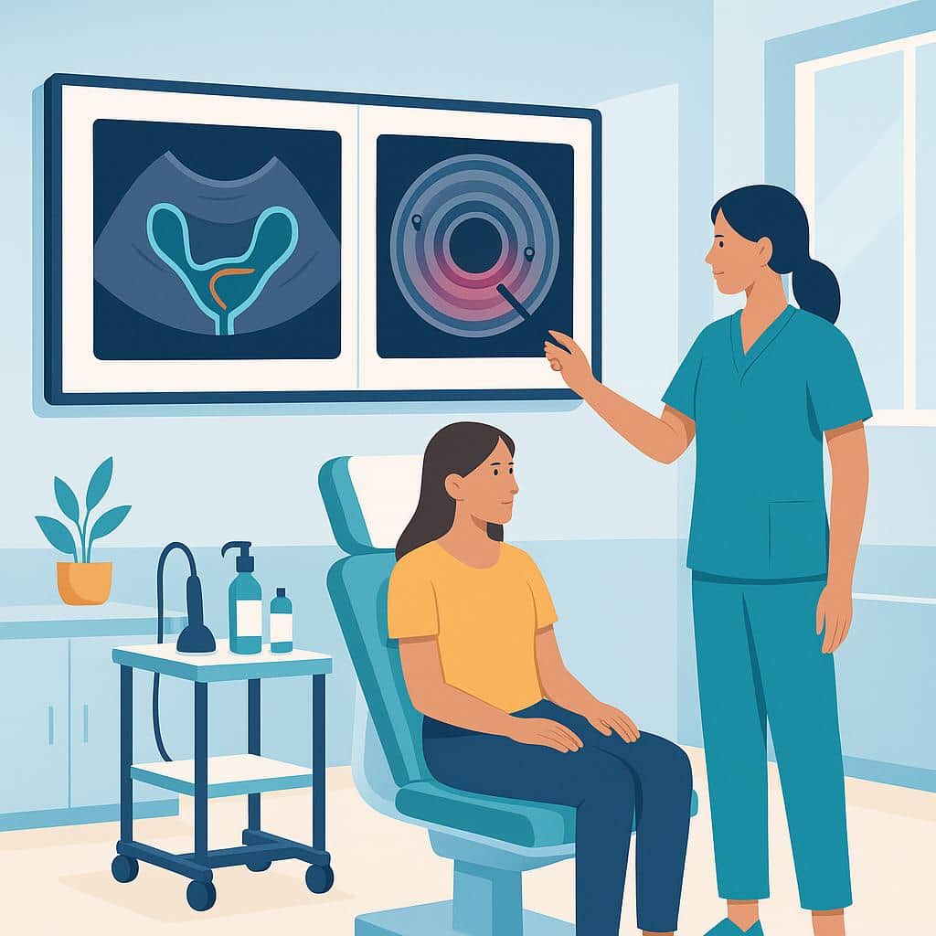
Many surgeons prefer endometriosis imaging within about 3–6 months of surgery, and sooner if your symptoms change or escalate. A radiologist or sonographer with specific endometriosis experience matters just as much as timing, because technique and interpretation can affect what is seen.
In most cases, we prefer relatively recent imaging so it reflects your current anatomy and helps with surgical planning. Many surgeons prefer imaging done within about 3–6 months of the operation, and sooner if your symptoms change or escalate.
Who performs the study matters just as much as timing. We typically recommend a radiologist or sonographer with specific endometriosis experience because technique and interpretation can affect what is and isn’t seen. Our team can help you choose the right type of imaging for your symptoms and ensure the images and report are in a format we can review efficiently before surgery. Reach out to schedule a consultation if you’d like guidance.
Where to go from here
Understand it, compare it with what you're feeling, then talk it through with us.
Compare
Symptoms it can relate to
Speak with Us
Get Accurate Imaging Timing
Recent imaging is crucial for effective surgical planning. Our specialists ensure your scans are done right to match your current symptoms and needs.
Questions people ask next
All diagnosis questionsDo I need imaging or special prep before endometriosis excision?
In many cases, yes: targeted transvaginal ultrasound and/or pelvic MRI can help plan endometriosis excision more safely and thoroughly. Prep depends on imaging and symptoms; some patients may need bowel preparation or temporary ureteral stents.
Ultrasound or MRI before endometriosis excision: which is best?
Neither ultrasound nor MRI is universally best before endometriosis excision. Expert ultrasound assesses deep disease and organ mobility; MRI often adds a broader map of disease near the bowel, bladder and ureters, and both may be recommended in complex cases.
Can imaging be negative and still help plan endometriosis surgery?
Yes, imaging can be negative and still help plan endometriosis surgery. Small or superficial disease may not show up, but imaging can help map deeper disease and assess whether nearby organs may be involved, helping plan the surgical team and anticipate complexity.
When is imaging most helpful for endometriosis?
Imaging is most helpful when clarifying what may be driving your symptoms before treatment, or when we suspect deeper disease that may involve the bowel or bladder. It can help guide surgical planning, but it can’t rule out endometriosis.
Further reading
Do You Need Two Ultrasounds Before Bowel Endometriosis Surgery?
How TVS (transvaginal) and ERUS (endorectal) map rectal endometriosis, guide bowel surgery planning, flag stenosis and risks, and who benefits.
Getting Ready for Endometriosis Surgery: A Week‑by‑Week Guide
Get ready for your endometriosis surgery with our week-by-week guide. Learn what to schedule, pack, and prepare for a smooth recovery.
What to Expect Before, During, and After Endometriosis Surgery
Discover what to expect before, during, and after endometriosis surgery. Insights on pain relief, preparation, recovery from reliable recent research.